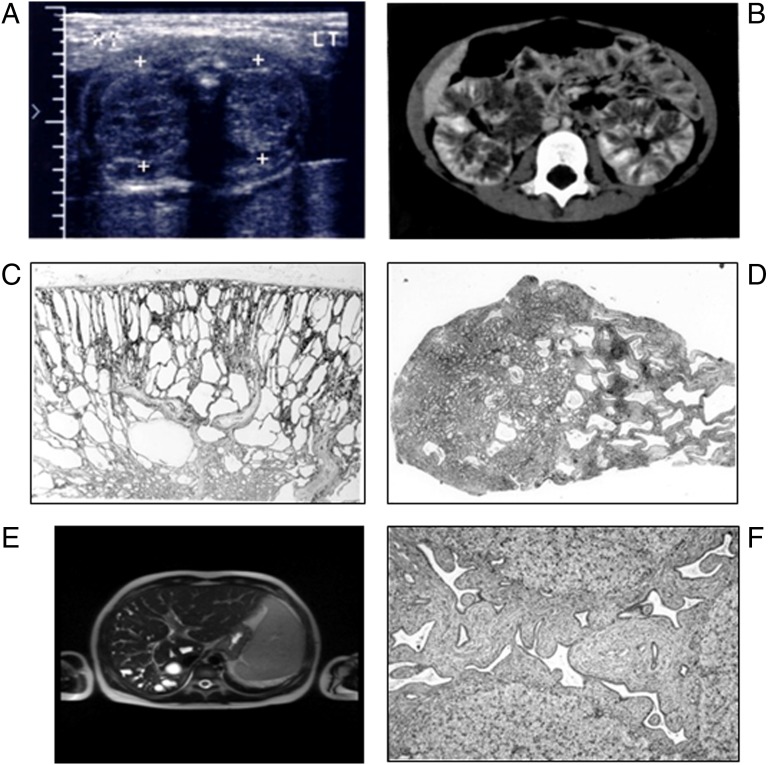
FIGURE 2.
Radiologic findings and pathologic features associated with ARPKD. A, Neonatal sonography with nephromegaly and increased echogenicity. B, Contrast-enhanced computed tomography in a symptomatic 4-year-old girl reveals a striated nephrogram and prolonged corticomedullary contrast retention. C, Light microscopy: ARPKD kidney from a 1-year-old child reveals discrete medullary cysts and dilated collecting ducts, hematoxylin and eosin (H&E) ×10. D, Light microscopy: later-onset ARPKD kidney with prominent medullary ductal ectasia, H&E ×10. E, Coronal heavily T2-weighted image of the abdomen in an 8-year-old boy reveals marked cystic and fusiform dilatation of the intrahepatic biliary system. F, Light microscopy: congenital hepatic fibrosis with extensive fibrosis of the portal area; ectatic, tortuous bile ducts; and hypoplasia of the portal vein, H&E ×40.

