
Fig. 2. Calcium oxalate stones (arrowheads) in 46-year-old man with history of gastric bypass, hyperoxaluria, and recurrent stones.
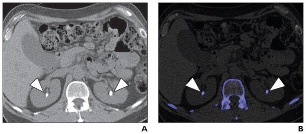
A, Axial unenhanced image of mid kidneys show bilateral renal calculi.
B, Axial unenhanced image of mid kidneys show bilateral renal calculi (blue) color coded to represent non–uric acid stones.