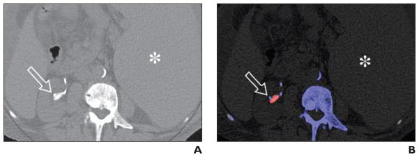
Fig. 3. Uric acid stones (arrow) in 54-year-old man with history of myelofibrosis and splenomegaly.

A, Axial unenhanced image of mid right kidney shows right renal pelvic stone and adjacent catheter tubing.
B, Axial unenhanced image of mid right kidney shows color-coded stone in mid right kidney. Red color coding is most consistent with uric acid composition. Note adjacent color-coded ureteral stent. Spleen (asterisk, A and B) is also enlarged.
