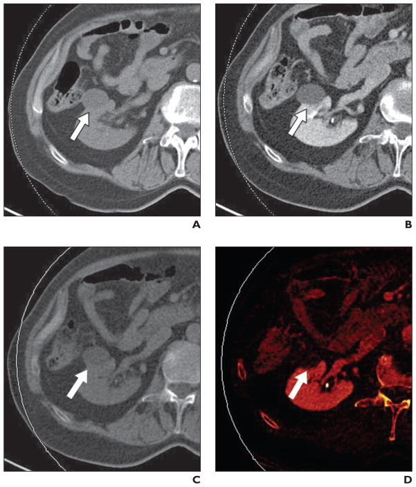
Fig. 4. Benign simple renal cyst (arrow) in 79-year-old man.
A, Axial unenhanced image through low-attenuation mass in right kidney. Attenuation of lesion measures 7 HU.
B, Axial enhanced image through lesion shows no visual evidence of enhancement. Attenuation of lesion measures 8 HU. Absence of enhancement is diagnostic of benign renal cyst.
C, Axial virtual noncontrast image. Measured attenuation was nearly identical to true unenhanced value (6.2 HU).
D, Axial iodine-overlay image through lesion shows absence of central iodine signal; this finding is consistent with diagnosis of renal cyst.

