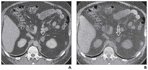
Fig. 6. Adrenal nodule (arrow) in 57-year-old man.

A, Axial contrast-enhanced image through nodule in left adrenal gland. Attenuation of lesion measures 48 HU.
B, Virtual noncontrast CT image through left adrenal mass created from contrast-enhanced images lesion shows –4 HU. Low attenuation changes of mass are consistent with benign adenoma; however, further research and clinical validation are required.
