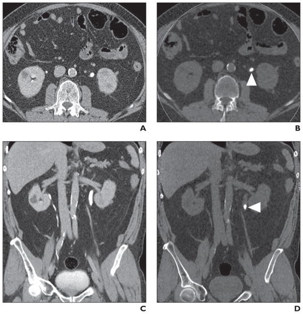
Fig. 7. Left ureteropelvic stone (arrowhead) in 65-year-old man visualized on virtual noncontrast image created from pyelographic phase.
A, Axial urographic phase image through mid kidneys with contrast material within both ureters.
B, Axial virtual noncontrast image shows left ureteropelvic stone.
C, Coronal urographic phase image through urinary tract with opacification of segments of collecting systems of both kidneys.
D, Coronal virtual noncontrast image shows calculus in proximal left ureter.

