Figure 3.
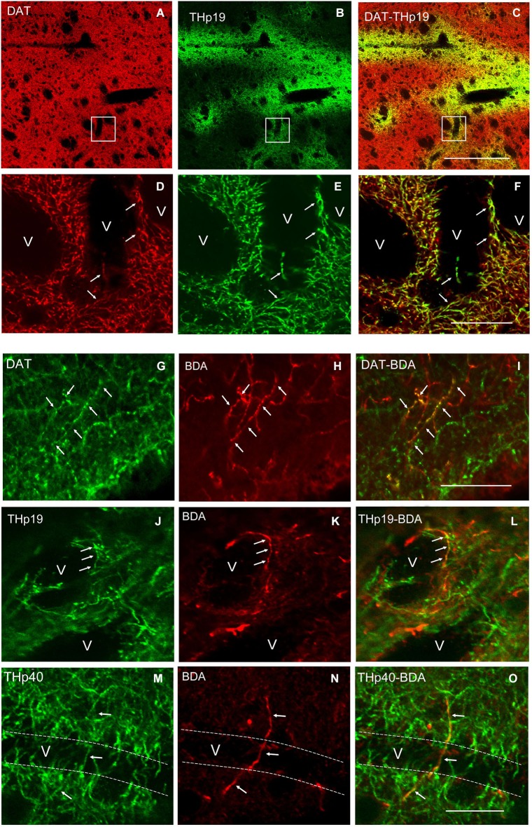
(A–C) A low magnification view of double labeling for the dopamine transporter (DAT) and THp19 in the dorsal striatum of the rat. (D–F) Boxed areas in (A), (B) and (C) respectively showing that all perivascular THp19 fibers and terminals are dopaminergic (DAT-immunoreactive). (G–O) Double labeling for DAT, THp19 or THp40 and BDA showing that BDA-positive ascending axons (H) and perivascular terminals (K,N) are immunoreactive for DAT (G–I), THp19 (J–L) and THp40 (M–O). v, vessel lumen. Dotted lines in (M–O) indicate the probable localization of the vessel wall. Bar in (C) (for A–C), 130 μm; in (F) (for D–F), 40 μm; in (I) (for G–I), 40 μm; in (O) (for J–O), 40 μm.

