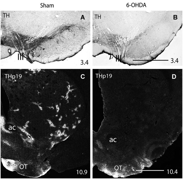
Figure 4.

(A,B) Immunohistochemistry for TH in the rat midbrain after sham (A) and 400 μg 6-OHDA (B) intracerebroventricular injection. (C,D) Immunofluorescence for THp19 in the striatum of sham (C) and 400 μg 6-OHDA (D) injected rats. One can see the decrease in the number of TH-positive midbrain cells after 6-OHDA injection (compare A and B), and that THp-19 striatal perivascular plexuses disappear after midbrain DA-cell degeneration (compare C and D). ac, anterior commissure; OT, olfactory tubercle; III, fibers of the third cranial nerve emerging in the surface of the ventral midbrain. Numbers at the bottom right indicate the distance from the interaural axis in millimeters. Bar in (B) (for A and B), 1 mm; in (D) (for C and D), 1 mm.
