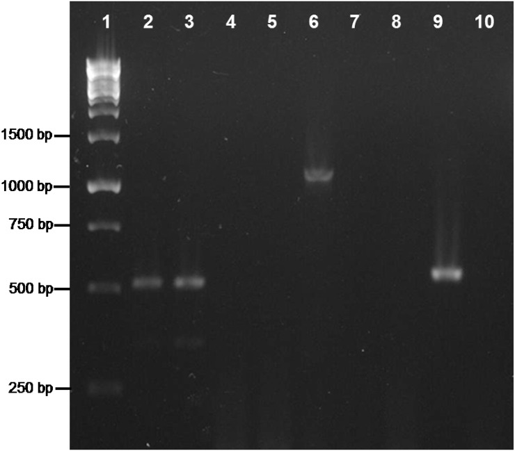
Figure 5.
PCR analysis on TDNA extracted from P. peruvianus. Heads (lines 2, 5, 8) and bodies (lines 3, 6, 9) were analyzed. Lane 1, Molecular Weight Marker; lines 2–4, amplification of 18S rDNA; lanes 5–7, amplification of the 16S rDNA; lanes 8–10, amplification of trpB. Negative controls: lanes 4, 7, 10.

