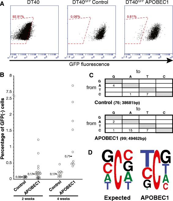
Figure 1.

APOBEC1 induces mutations in DT40 cells. (A) The inactivation of the EGFP transgene in DT40 cells was assayed by monitoring the size of a distinct EGFP(−) population in transfectants expressing either APOBEC1 or a control plasmid (Figure S1A in Additional file 1). Flow cytometric analysis of representative DT40GFP clones stably transfected with APOBEC1 or with the control plasmid. The red boxed area indicates the EGFP(−) population considered for the analysis. (B) Plot depicting the inactivation of the EGFP in independent transfectants, with the median for each construct indicated. The measurements were taken at the indicated times (P < 0.05 for each time-point). The sorted EGFP(−) cells did not regain fluorescence after expansion. (C) Mutation pattern obtained after sequence analysis of independent EGFP fragments from sorted EGFP(−) APOBEC1- and vector-transfected DT40 cells. The number of sequences analyzed and the total number of bases is indicated in parentheses (P = 0.012 by Fisher’s exact test). Only non-clonal mutations were considered, which is likely to underestimate the number of mutations in the case of preferred mutational hotspots. (D) Local sequence context for the cytosine residues present on both strands of the analyzed GFP fragments (expected) and for the mutated cytosines (observed) in the APOBEC1-expressing clones.
