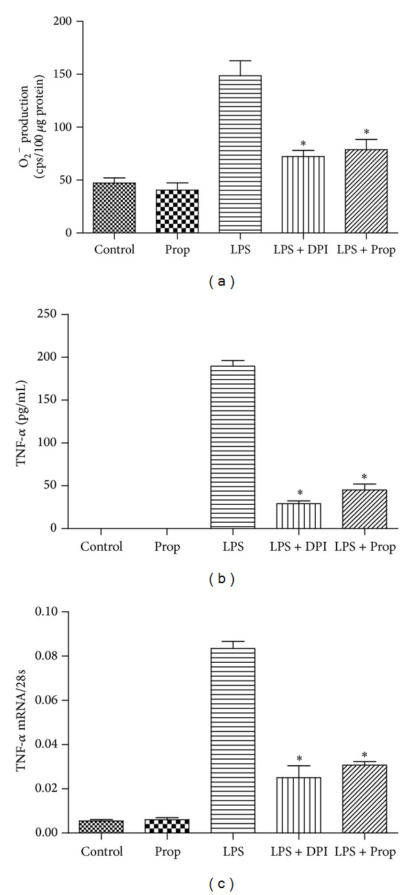
Figure 3.

The inhibitory effect of propofol on O2 − generation and TNF-α expression in LPS-stimulated cardiomyocytes. Cardiomyocytes were pretreated with vehicle, DPI (50 μM), or propofol (50 μM) followed by LPS (4 μg/mL) for 2 or 4 hours. (a) O2 − generation was determined by lucigenin-enhanced chemiluminescence at 2 hours after LPS treatment; (b) TNF-α protein measurement by ELISA at 4 hours after LPS treatment; (c) TNF-α mRNA measurement by real-time RT-PCR at 2 hours after LPS treatment (each bar represents the mean ± S.D, *P < 0.05, compared with LPS group; n = 4).
