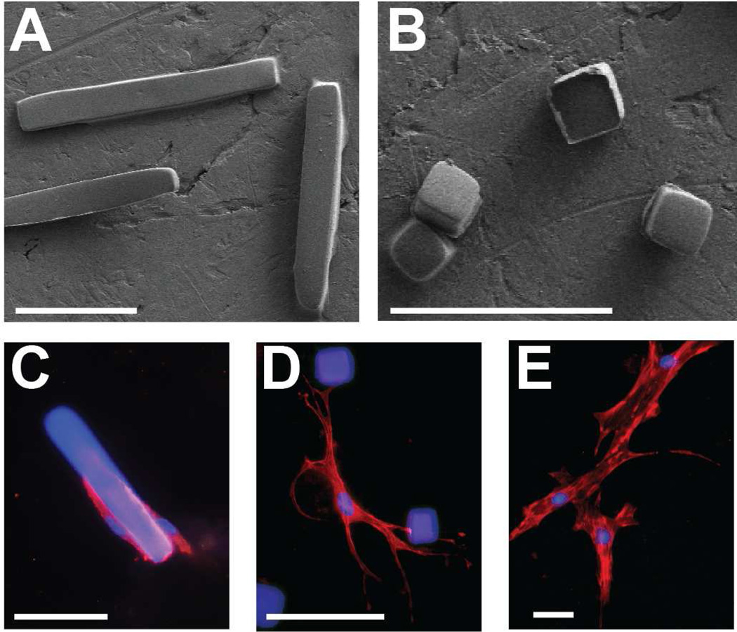
Fig. 2. Microrods and microcubes interact with fibroblasts in vitro.
Scanning electron micrograph of free-standing microstructures fabricated from polymerized polyethylene glycol dimethacrylate (“microrods”, 100 µm × 15 µm × 15 µm (A); “microcubes”, 15 µm × 15 µm × 15 µm (B)). Fluorescent immunocytochemical stain of adult Sprague-Dawley ventricular cardiac fibroblasts interacting with a microrod (C), microcubes (D), or in the absence of microstructures (E) suspended in a 3D culture of Matrigel. Scale bar = 50 µm (red = rhodamine phalloidin, blue = nuclei and microstructures).

