
Figure 1).
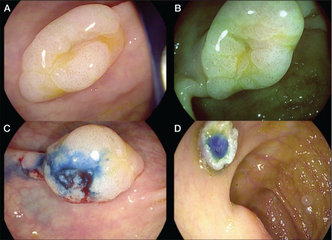
A High-definition iSCAN (Pentax, Japan) technique showing a flat lesion in the ascending colon. B and C iSCAN virtual chromoendoscopy characterized the margins of the lesion and the Kudo pit pattern of the mucosa as type II open and irregular spiral vascular pattern. D Endoscopic mucosal resection after previous injection of saline and methylene blue was performed. Serrated adenoma was reported by histology