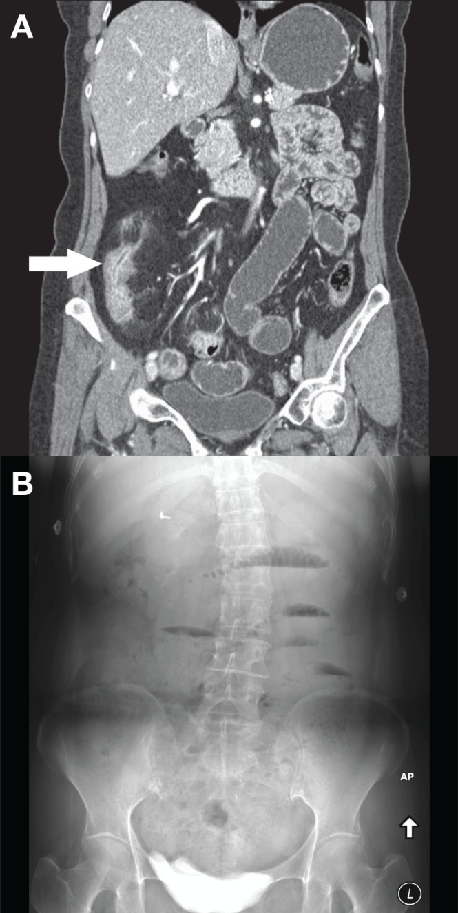
Figure 1).

A Coronal computed tomography enterography image demonstrating active inflammation of the terminal ileum (arrow). The upstream small bowel is filled with oral contrast. B Upright abdominal radiograph performed 9 h later demonstrating dilated small bowel with air-fluid levels consist with a small bowel obstruction
