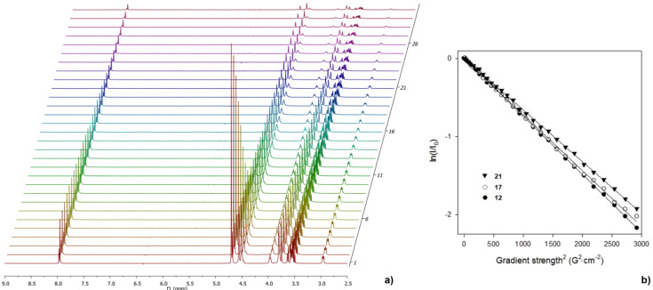
Figure 1.
a) Decay of 1H signal for the nonavalent mannosylated compound 12 in D2O during the PFGSTE experiment. The gradient strength is increased linearly between 1.8 and 54.2 G·cm−1; b) characteristic echo decays of the H-5 resonances (δ = 2.98 ppm) as a function of squared gradient strength located in 12 (full circles) and 21 (full triangles) with δ = 4 ms and Δ = 50 ms (Δ = 40 ms for 17 (circles)). Notably, such linear behavior was also obtained for the decay of the signal intensities of other protons located either in internal regions of the conjugates on aromatic or branching sections, or in the peripheral saccharidic belt (results not shown).

