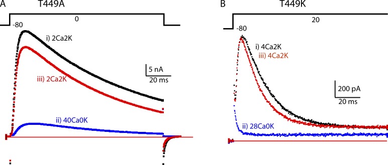
Figure 1.
In two mutants that have C-type inactivation, high extracellular Ca2+ depresses IK. (A) The traces show IK from a cell expressing T449A during a depolarization lasting 110 ms from −80 to 0 mV. The traces were taken in the order i–iii, with a rest of ∼2 min between them to allow full recovery from C-type inactivation. IK is strongly depressed by 40Ca0K. Note, however, that the time constant of inactivation of the current remaining in 40Ca0K is similar to that in 2Ca2K. (B) IK traces from a T449K cell. The extracellular solution was changed from 4Ca2K (i) to 28Ca0K (ii) and then back to 4Ca2K (iii).

