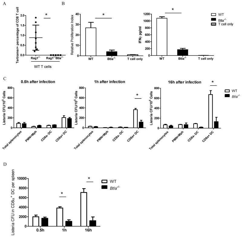
Figure 4.
BTLA promotes proliferation of Listeria in CD8α+ DC s for better CD8+ T cell responses. A) Rag1−/− and Rag1−/− Btla−/− mice were adoptively transferred with 2 × 10⁷ splenocytes. Fourteen days later, mice are infected with Listeria-OVA and OVA specific T cell responses were detect 7 days after infection. B) WT and Btla−/− mice were infected i.v. with 1×10⁷ Listeria-OVA. Sixteen hours after infection, sorted splenic CD8α+ CD11c+ DCs were incubated with purified OVA-specific OT -I T cell. 3H-thymidine was pulsed in the last 18 hours of a three-day culture and cells were harvested for liquid scintillation counting. IFN-γ production in the supernatant was measured on day 2 of the culture. C) WT and Btla−/− mice were infected i.v. with 1×10⁷ Listeria -OVA. Thirty minutes, 1 hour and 16 hours after infection, splenic CD11cloCD8αloCD11b+ granulocyte (PMN) and macrophage (Mφ, including monocytes), CD8α+ and CD8α− CD11c+ DC populations were purified by flow sorting, and sorted populations were lysed and plated onto BHI agar to determine degree of infection. D) Total Listeria CFU in CD8α+ DC per spleen was shown from the same experiment as C). The results are representative of three independent experiments, three to five mice in each group and data are represented as mean +/− SEM. See also Figure S4.

