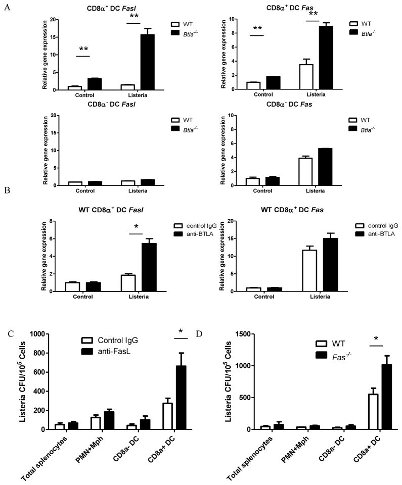
Figure 6.
BTLA inhibits the expression of FasL and Fas during Listeria infection. A) Sorted splenic CD8α+ and CD8α− DCs from WT and Btla−/− mice were infected with Listeria-OVA in vitro for 6 hours. The expression level of fasL and fas mRNA was analyzed by quantitative real-time PCR. B) Sorted splenic CD8α+ DCs from WT were infected with Listeria-OVA for 6 hours in the presence of hamster Ig or anti-BTLA (6A6) Ab. The expression level of fasL and fas mRNA was analyzed by quantitative real-time PCR. The results are representative of three independent experiments. C) Btla−/− mice were infected i.v. with 1×10⁷ Listeria -OVA and treated with hamster Ig or anti-FasL Ab. Sixteen hours after infection, splenic CD11cloCD8αloCD11b+ granulocyte (PMN) and macrophage (Mφ, including monocytes), CD8α+ and CD8α− CD11c+ DC populations were purified by flow sorting, and sorted populations were lysed and plated onto BHI agar to determine degree of infection. D) WT and Fas−/− mice were infected i.v. with 1×10⁷ Listeria-OVA. Sixteen hours after infection, splenic CD11cloCD8αloCD11b+ granulocyte (PMN) and macrophage (Mφ, including monocytes), CD8α+ and CD8α− CD11c+ DC populations were purified by flow sorting, and sorted populations were lysed and plated onto BHI agar to determine degree of infection. The results are representative of two independent experiments, four mice in each group and data are represented as mean +/-SEM. See also Figure S5.

