Figure 3.
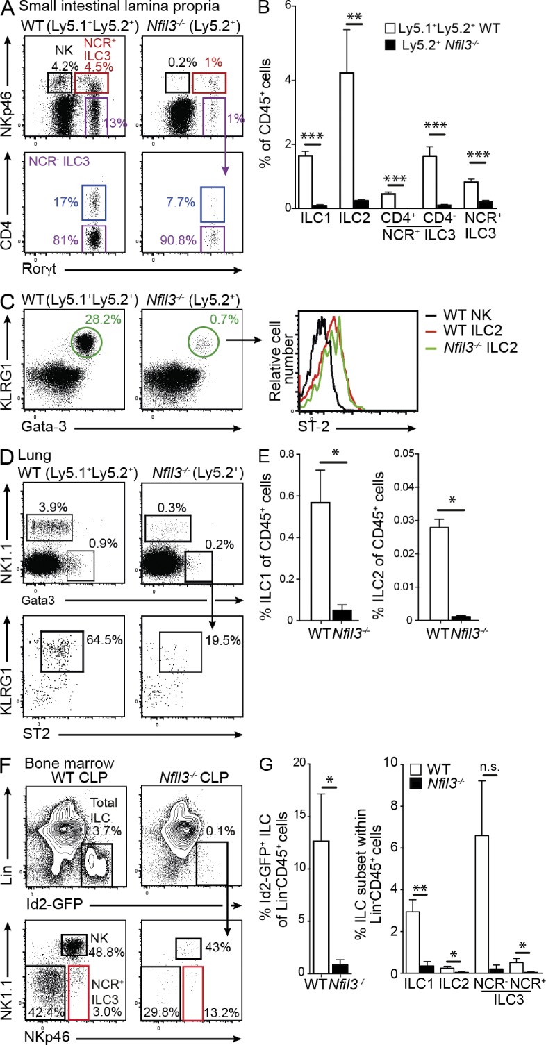
Nfil3 regulation of ILC development is cell intrinsic. Lethally irradiated WT (Ly5.1+) recipient mice were reconstituted with an equal mix of WT (Ly5.1+Ly5.2+) and Nfil3−/− (Ly5.2+) bone marrow cells. After 8 wk, the proportion of ILC1, ILC2, and ILC3 were determined in the small intestinal lamina propria (A–C) and the lung (D and E). (A and C) Dot plots show representative profiles of ILC3 (A) and ILC2 (C, left) in Nfil3-sufficient and -deficient compartments gated on Lin− (CD3−CD19−) CD45+ hematopoietic cells. (B) Frequency of ILC subsets within the WT (Ly5.1+Ly5.2+) and Nfil3−/− (Ly5.2+/+) populations in mixed chimeric mice. Data show the mean ± SEM (n = 3 mice/genotype) of one representative of two experiments. Statistical differences were tested using an unpaired student’s t test. (C) Expression of ST-2 within the KLRG1+Gata-3+ ILC2 subset (right). (D) Expression of NK1.1 and Gata-3 in Lin− (CD3−CD19−Gr1−) CD45+ cells isolated from the lungs and KLRG1 and ST2 expression on ILC2 (bottom). (E) Frequency of the different ILC1 and ILC2 in lung in each WT (Nfil3+/+) and Nfil3−/− compartment in mixed chimeras. (A–E) Analyses show representative profiles of two experiments with 6 mice/genotype. (F and G) Flow cytometrically purified CLP (Lin−CD127+Flt3/Flk2+Sca1intCD117int) isolated from bone marrow of Id2gfp/gfp mice were adoptively transferred into Rag2γc−/− mice. After 2 wk, the reconstitution of ILC subsets in the small intestinal lamina propria was analyzed. Representative flow cytometric plots show the proportion of total Id2-GFP+CD45+Lin− (CD3−CD19−CD11c−Gr1−) ILC (top) and ILC subsets within that gate (bottom) in WT and Nfil3−/− mice. (F) Data show representative flow cytometric profiles from (G) individuals pooled from two experiments and show mean ± SEM (n = 4 mice/genotype). Statistical differences were tested using an unpaired Student’s t test. *, P < 0.05; **, P < 0.01; ***, P < 0.001; n.s., not significant.

