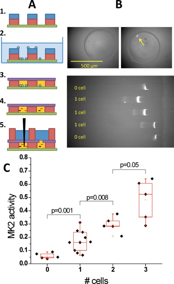
Figure 3.

Demonstration of single cell concentration-enhanced kinase activity assay. (a) Procedures for single cell culture, lysis and kinase reaction. Individual cells are isolated and cultured in individual open microwells (∼40 nL) for microscopic observation. For kinase assay, buffer in the wells is replaced with kinase assay buffer, sealed with a Kapton tape, and the cells are ultrasonically lysed. After incubation, to allow kinase reaction, the reaction product is diluted into larger volumes and loaded into the microflluidic device for readout. (b) Detection of 0 vs 1 cell Akt activities from different microwells. Measured activity signals (marked by arrows) can be clearly differentiated between 0 and 1 cell cases. (c) MK2 activity vs cell number after normalizing by the average cell volume. Different numbers of cells have significantly different normalized kinase activities by Student’s two-tailed t-test, demonstrating that this assay has both single cell sensitivity and resolution.
