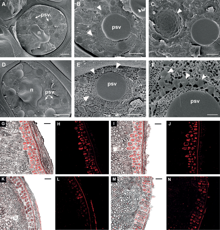
Fig. 2.
Oil body morphology in barley grains. (A–F) SEM cryo-fractured images of wild-type (var. ‘Golden Promise’) barley aleurone cells. (A–C) Two day imbibed grains; (D, E), 5 d imbibed grains. (A, D) Aleurone cells, showing nucleus (n) and numerous protein storage vacuoles (psv). (B, E) PSV, surrounded by oil bodies (OBs), indicated by arrowheads. (C) PSV fractured across, surrounded by OBs; the arrowhead indicates the extracellular face (‘E-face’) where PSV and OBs have been removed during fracture. (F) Close-up of (E), showing connections between OBs and PSV. OBs are indicated by arrowheads. (G–N) Confocal images of unfixed barely grain sections showing Nile Red staining of OBs in aleurone cells. De-embryonated grains were imbibed for 24h (G–J) or 96h (K–N) in the presence of 1 μM GA. (G, H, K, L) HvABCD1/2 RNAi line 5; (I, J, M, N) null segregant. In G, I, K and M, the confocal image is overlaid on the bright field image. Scale bars: A=5 μm, B, C, E=1 μm; D=4 μm; F=400nm; G–N=50 μm.

