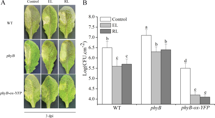
Fig. 1.
Effect of exposure to EL and RL on disease progression in leaves of WT, phyB, and phyB-ox-YFP plants. (A) After exposure to EL (1500 μmol photons m–2 s–1for 1h), and excess RL (120 μmol photons m–2 s–1 for 4h), WT and phyB, phyB-ox-YFP plants were inoculated with virulent Pst-DC3000 (OD600=0.01 in 10mM MgCl2). Leaves were infected on their left halves, and samples were collected at 3 d post-inoculation (dpi). (B) Bacterial growth quantification of Pst-DC3000-inoculated (OD600=0.0001) leaves after exposure to EL and RL. Samples were collected at 3 dpi for the assay. Each value is the mean±standard deviation (SD) of three replicates. Different letters indicate statistically significant differences between treatments (Duncan’s multiple range test: P<0.05). CFU, colony-forming units. (This figure is available in colour at JXB online.)

