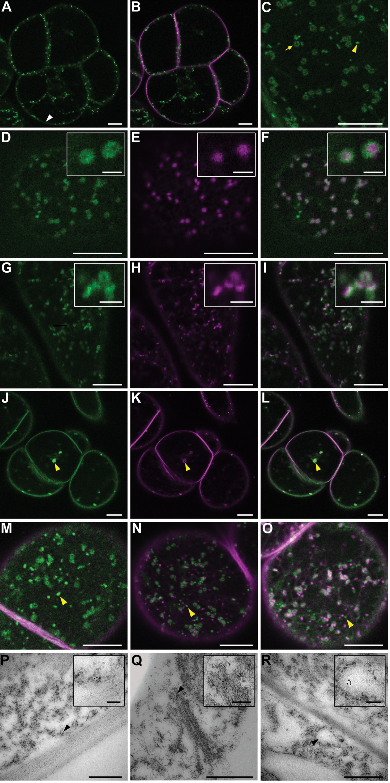
Fig. 2.
RBOHDs mostly localize to the PM and Golgi in BY-2 cells, as shown by confocal microscopy of RBOHD1-GFP-expressing BY-2 cells (A–O) and immunogold labelling of RBOHDs on ultrathin sections of wild-type cells (P–R). (A–C) Cells expressing RBOHD1-GFP and stained with the endocytic tracer FM4-64 for 5min. Shown are GFP fluorescence alone (A, C) and an overlay of GFP and FM4-64 fluorescence (B). GFP signals were observed at the PM [white arrowhead in (A)] and in intracellular dots and rings [yellow arrowhead and yellow arrow, respectively, in (C)]. (D–F) Cells co-expressing RBOHD1-GFP and the Golgi marker Man99-mRFP. Shown are GFP fluorescence (D), mRFP fluorescence (E), and overlay (F). The insets show that RBOHD1-GFP labelled the margin of the Golgi. (G–I) Cells expressing RBOHD1-GFP and stained with the endocytic tracer FM4-64 for 30min. Shown are GFP fluorescence (G), FM4-64 fluorescence (H), and overlay (I). The inset in (I) shows tricoloured labelling due to partial overlap. (J–L) Cells expressing RBOHD1-GFP, treated with BFA for 60min and with FM4-64 for 30min. Shown are GFP fluorescence (J), FM4-64 fluorescence (K), and overlay (L). The yellow arrowhead indicates a BFA body. (M–O) RBOHD1-GFP-expressing cells were stained with FM4-64 for 10min (M), 20min (N), and 60min (O). Shown are overlays of GFP and FM4-64 fluorescence. Yellow arrow and arrowheads indicate FM4-64-labelled tonoplast and green-only dots, respectively. (P–R) Immunogold-labelled sections of BY-2 cells performed with anti-RBOHD antibody. Black arrowheads indicate GPs associated with the PM (P), the margin of the Golgi (Q), and a vesicle-like compartment (R). Scale bar represents 10 µm (A–O), 2 µm (insets in panels D–I), 500nm (P–R), and 100nm (insets in panels P–R).

