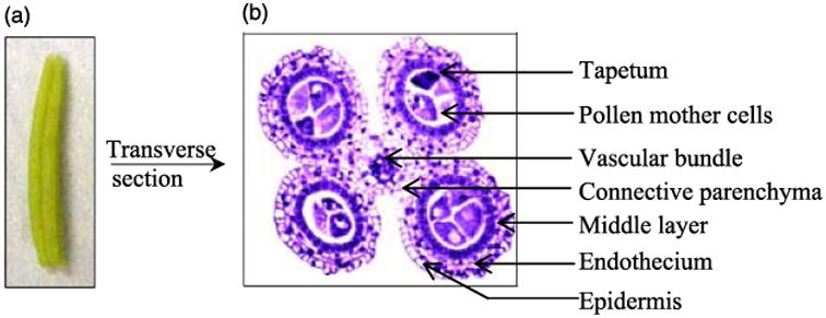
Figure 1.

Maize anther morphology and anatomy. (a) Photograph of a 1.5-mm maize anther when the PMC is ready for meiosis; (b) In transverse view (safranine and fast green staining, Wang et al., 2009), each anther has four locules, a single vascular bundle and surrounding connective parenchyma. The anther lobe somatic tissue is composed of four wall layers: epidermis, endothecium, middle layer and tapetum. PMC is located centrally. Meiosis, from the 1.5–2.5-mm anther stages, results in four microspores from each PMC. These separate from the tetrad and mature into pollen.
