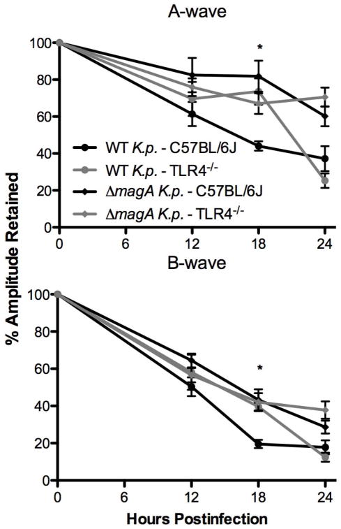
Figure 2. Evaluation of Retinal Function.
After infection with K. pneumoniae, mice were dark adapted for at least 6 hours. At the indicated time points, mice were anesthetized as described above and subjected to electroretinography. The average of n>6 eyes ± SEM is shown for each time point. *;p≤0.01 for C57BL/6J –wild type K. pneumoniae. vs. TLR4−/−–wild type K. pneumoniae

