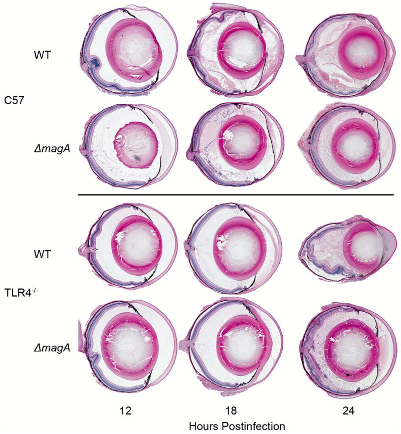
Figure 3. Ocular Histology of C57BL/6J and TLR4−/− Mice during Experimental K. pneumoniae Endophthalmitis.
At the indicated time points postinfection, mice were euthanized and whole globes were harvested, fixed, sectioned and stained with hematoxylin and eosin as described above. Each eye is representative of an n≥3, except the 24 hour time point for wild type K. pneumoniae in C57BL/6J mice. At this time point only one eye had not undergone pthisis and could be removed intact.

