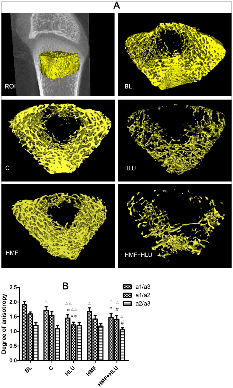
Figure 5. Effects of HMF and HMF+HLU on the femur trabecular bone.
ROI: A 2.019-mm-thick trabecular bone chip under the epiphyseal plate in the lower end of the femur was selected as the region of interest (ROI). BL: The baseline group. Rats were executed to get basal data on day 0 of the experiment. C: Rats were raised in a wooden box with a normal GMF for 28 days; HLU: Rats were suspended, unloaded with −30° downward head tilting, and raised in a wooden box; HMF: Rats were raised normally in a GMF-shielded room; HMF+HLU: HLU rats were raised in a GMF-shielded room. (A) Three-dimensional trabecular bone architecture of an ROI located under the epiphysis plate of the femur. (B) The degree of anisotropy is a measure of the extent of the orientation of the substructures in an ROI of trabecular bone, which represents the directivity and symmetry of the trabecular structure; a1/a3, a1/a2, and a2/a3 are the ratios of the long to the short diameters on 3 mutually perpendicular plane ellipses inside the ellipsoid. **P<0.01 vs. C, *P<0.05 vs. C, # P<0.05 vs. HLU, △△ P<0.01 vs. BL, △ P<0.05 vs. BL.

