Fig. 5.

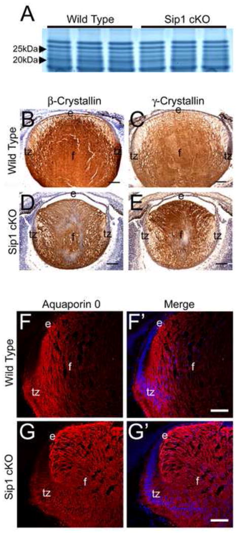
Analysis of fiber cell marker expression in Sip1 cKO lenses shows little to no change in embryonic crystallin expression. SDS-PAGE analysis shows little to no difference in global crystallin expression at E16.5 (A). Expression of β-crystallins (B) and γ-crystallins (C) in E16.5 sections, also appears normal in the Sip1 cKOs (D and E). Lastly, expression of Aquaporin 0, staining the fiber cell membrane in the wild type (F), also shows little difference in the Sip1 cKOs (G), although the staining is different reflecting the observed fiber cell tip migration defect (Prime panels (e.g. F′) shows Aquaporin 0 expression (Red) merged with nuclei (DRAQ5, Blue)). Abbreviations: e, lens epithelium; f, lens fiber cells; tz, lens transition zone. Scale Bars = 100μm (B – E); 77μm (F, G).
