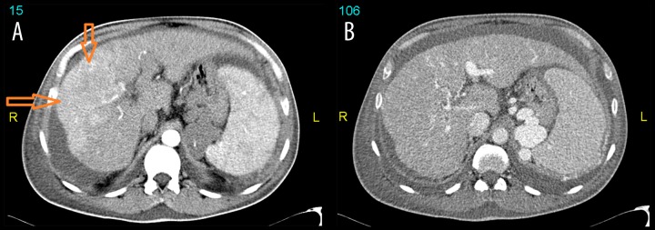
Figure 2.

CT scan of the upper abdomen in arterial (A) and venous (B) phases revealed a small cirrhotic liver with wide spread infiltrative type of hepatocellular carcinoma involving almost the whole right lobe and segment IV of the left lobe which showed patchy areas of arterial enhancement (arrows) with washout in venous phase. Splenomegaly and portal hypertension associated with spleno-renal shunt and extensive varicose veins formation is seen as well as bilateral pleural effusion and ascites.
