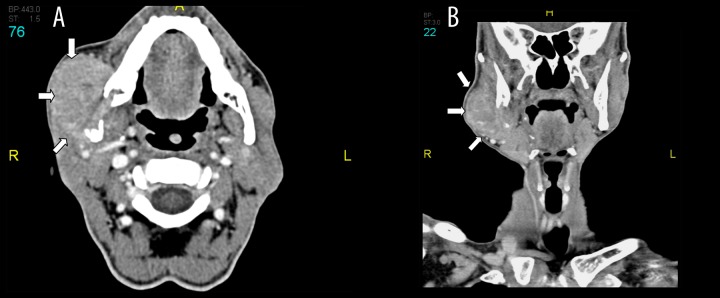
Figure 3.

Post contrast CT scan axial (A) and coronal (B) shows right mandibular angle osteolytic destructive lesion with buckle side soft tissue mass infiltrating the masseter muscle and ventrally indenting the right parotid salivary gland (block arrows).
