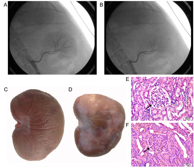
Figure 1. Images and morphological analysis of the left kidney.
Images of left renal artery angiography before transcatheter embolization (A) and after transcatheter embolization (B) in the model group. Small renal artery branches were occluded, whereas the main renal artery or sub-segment renal artery remained fluent in the model group. Gross appearance of the left kidney after 2 weeks of interventional operation in the sham group (C) and the model group (D). The left kidney in the model group became pale and had atrophy and infarction. Images of HE staining of the left kidney after 2 weeks of interventional operation in the sham (E) and model (F) groups. Glomeruli were severely damaged in the model group. Arrows show a glomerulus in the sham and model groups.

