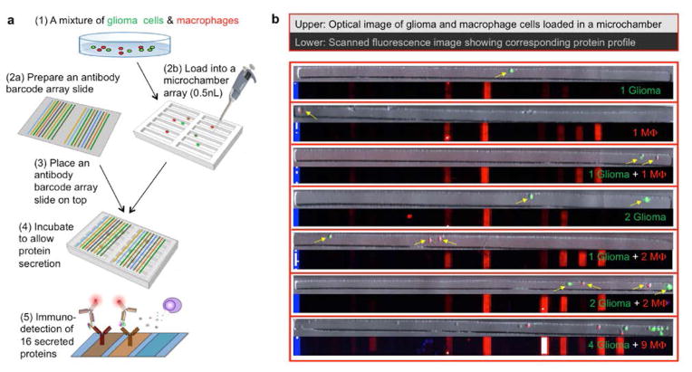
Figure 2. Microchip for protein secretion profiling of glioma and macrophage cells at the single-cell level.
(a) Work flow of high-throughput multiplexed single cell secretomic assay to measure glioma, macrophage and their combinations. (b) Optical micrograph of single macrophage (red), glioma (green) and glioma- macrophage co-culture microwells and their corresponding scanned fluorescence images showing the raw data of single cell secretomic measurements. Blue lines are position marks created with immobilized Fluorescein Isothiocyanate labeled bovine serum albumin (FITC–BSA, 488). Red is APC dye-streptavidin (Cy5, 635) for specific protein detection within each microwell.

