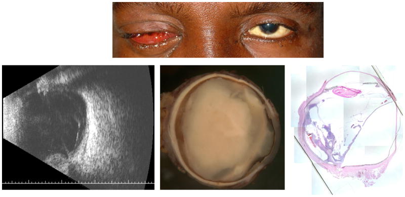
Figure 2.

Patient 1 was a 43-year-old man with no past medical history who presented with pain, chemosis, and finger count vision in the right eye (top). Posterior segment examination revealed dense vitritis with no view in the right eye. Initial B-scan ultrasonography confirmed no evidence of retinal detachment. The patient underwent vitreous tap and injection of intravitreal antibiotics. Vitreous culture grew Klebsiella pneumonaie. Systemic workup revealed positive blood cultures for Klebsiella pneumonaie and large liver abscess. The patient clinically worsened and repeat B-scan ultrasonography revealed a bullous retinal detachment and evidence of panophthalmitis (bottom left). The patient underwent enucleation one week after presentation (bottom center). Histopathlogic examination with hematoxylin and eosin at 1.25x magnification revealed a subretinal inflammatory collection underlying the detachment (bottom right).
