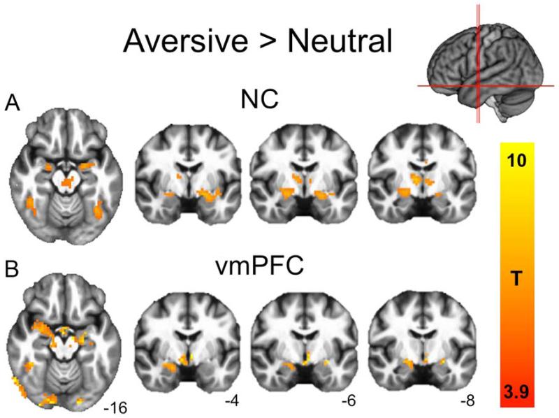
Figure 2.
Neural responses to aversive>neutral pictures. (a) NC subjects (PFWE<0.05; FWE, family wise error). (b) vmPFC lesion patients (displayed at corrected NC threshold of T=3.9 for comparison). Both groups exhibited robust bilateral amygdala responses, as well as responses in visual cortex, lateral temporal cortex, thalamus, and cingulate gyrus (see Table 2 for full cluster list).

