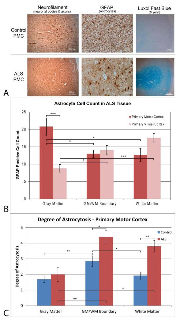
Figure 4.
– a) Histology stains, b) GFAP cell counts, and c) Degree of astrocytosis in ALS and control tissue samples. a) Antibody staining for neurons in gray matter (left – neurofilament), astrocytes in white matter (right - GFAP) and myelin (right - Luxol Fast blue) for control (top) and ALS (bottom) primary motor cortex tissue sections. Images were taken at 50x, 200x, and ~10x for the neurofilament, GFAP and Luxol stains, respectively, with scale bars at 200 μm, 50 μm, and 1mm. Neurofilament staining indicate that there is a decrease in neuronal body staining and reduction in protruding axons in ALS tissue. Astrocyte staining shows less proliferation of GFAP positive astrocytes in control tissue compared to ALS tissue. Astrocytes found in the ALS PMC show an upregulation of GFAP positive filaments indicative of a reactive state. Luxol fast blue staining of myelin is more pronounced in white matter of ALS PMC tissue, indicative of oligodendrocyte myelin sheath deterioration. b) GFAP cell counts of the ALS primary motor and primary visual cortices. A significant increase in GFAP positive astrocytes is found within the gray matter of the ALS PMC compared to ALS primary visual cortex internal control tissue as well as white matter and white/gray boundary ALS PMC tissue. c) Degree of astrocytosis for GFAP positive astrocytes in control and ALS tissue samples in the primary motor cortex. ALS PMC tissue exhibited significantly more astrocytosis in white matter and the gray/white matter boundary than control tissue. This increase in reactive astrocytosis was not seen in the gray matter of the PMC. *, p < 0.05; ** p < 0.01;, *** p < 0.001; **** p < 0.0001.

