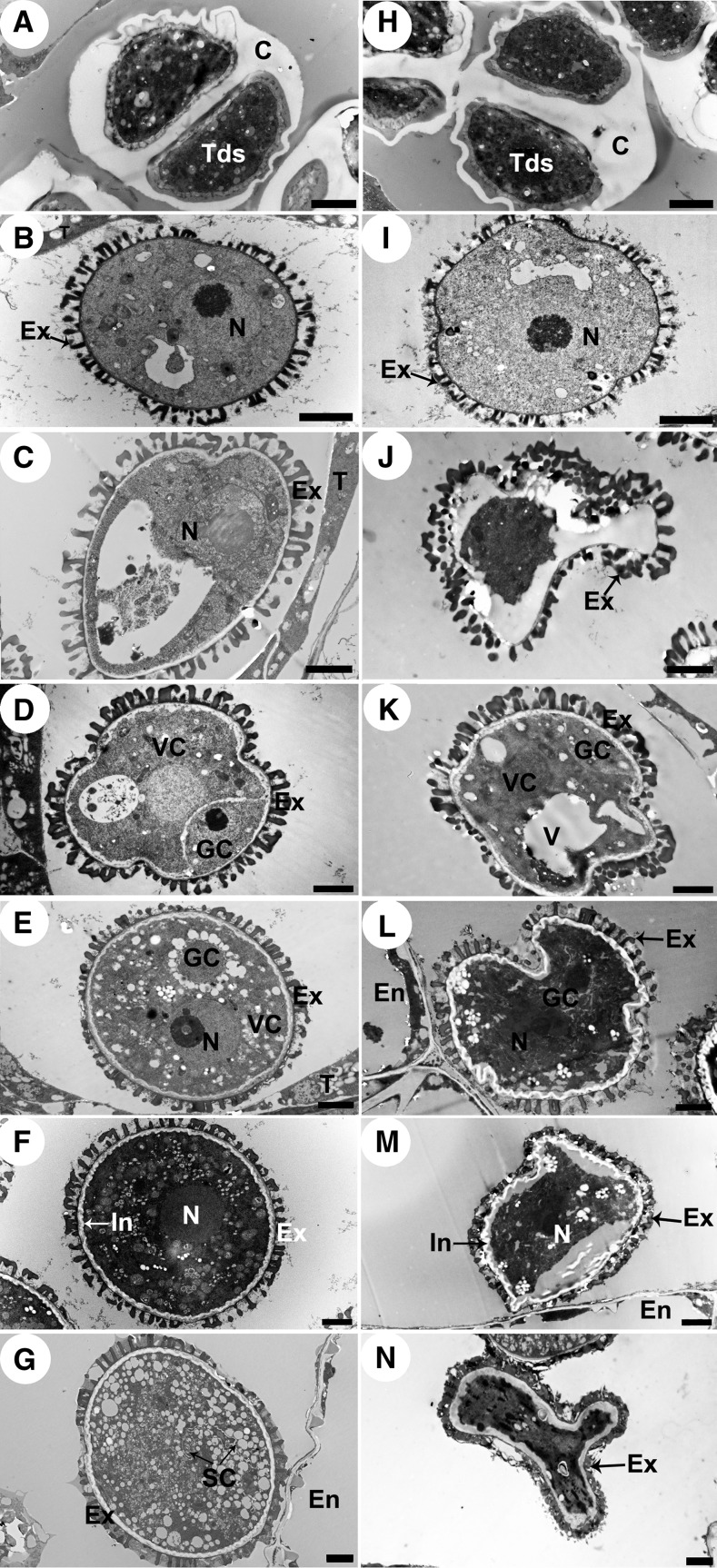
Figure 5.
Transmission Electron Micrographs of Microspores from the Wild Type and cep1 Mutant.
Microspores of the six developmental stages in the wild type ([A] to [G]) and cep1 mutant ([H] to [N]). (A) and (H), stage 7; (B) and (I), stage 8; (C) and (J), stage 9; (D) and (K), stage 10; (E) and (L), early stage 11; (F) and (M), late stage 11; (G) and (N), stage 12. C, callose; En, endothecium; Ex, exine; GC, generative cell; In, intine; N, nucleus; SC, sperm cell; T, tapetal cell; Tds, tetrads; V, vacuole; VC, vegetative cell. Bars = 2 μm.

