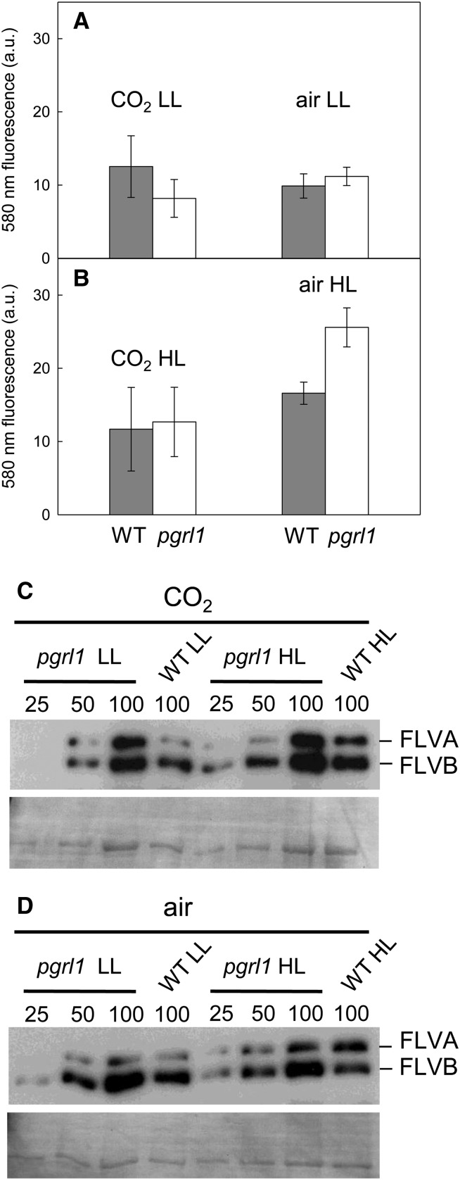
Figure 6.
Production of Extracellular Hydrogen Peroxide and Accumulation of FLVs by Wild-Type and pgrl1 Lines Grown under Different Light and CO2 Conditions.
(A) and (B) Measurements were performed on cells grown photoautotrophically in batch cultures supplied with 2% CO2 in air or with air in the presence of LL (50 μmol photons m−2 s−1) or HL (200 μmol photons m−2 s−1). Hydrogen peroxide concentration was assessed in culture medium using Amplex red by measuring fluorescence emission at 580 nm. Wild-type progenitor (dark bars) and pgrl1 (white bars) lines. Shown are means ± sd (n = 3).
(C) and (D) Accumulation of the FLV proteins was determined by immunoblot analysis of wild-type and pgrl1 lines grown in photobioreactors operated as turbidostats at a constant biomass concentration (≈1.5 × 10⁶ cells mL−1). The cultures were grown in 120 μmol photons m−2 s−1 (LL) or 500 μmol photons m−2 s−1 (HL) light intensity and in the presence of 2% CO2 in air (C) or in air (D). The FLVB protein has a size of ∼60 kD and FLVA shows the faint band at ∼70 kD. Fifteen micrograms of total protein as 100% was loaded per lane, and from the pgrl1 mutant 50 and 25% were loaded as well.

