Figure 1.
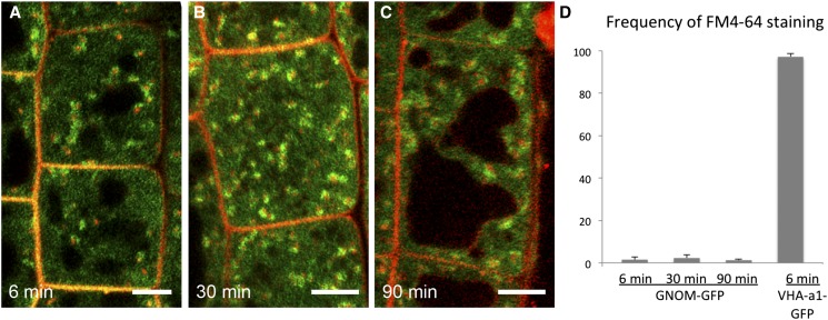
GNOM-GFP-Labeled Compartments Are Not Efficiently Stained by FM4-64.
(A) to (C) Representative confocal images of root epidermal cells labeled with FM4-64 (red) and GNOM-GFP (green). The time after the start of incubation with FM4-64 is indicated on each panel. Bars = 3 µm.
(D) Quantification of the colocalization ratio between GNOM-GFP and FM4-64. Most of the GNOM-GFP-labeled structures are not stained by FM4-64, even at a time when FM4-64 reaches the vacuoles, whereas the TGN/EE marker, VHA-a1-GFP, is strongly colocalized with FM4-64. Error bars indicate the sd values for data from three independent experiments. At least 200 GNOM-GFP-labeled organelles were analyzed in each experiment.

