Figure 4.
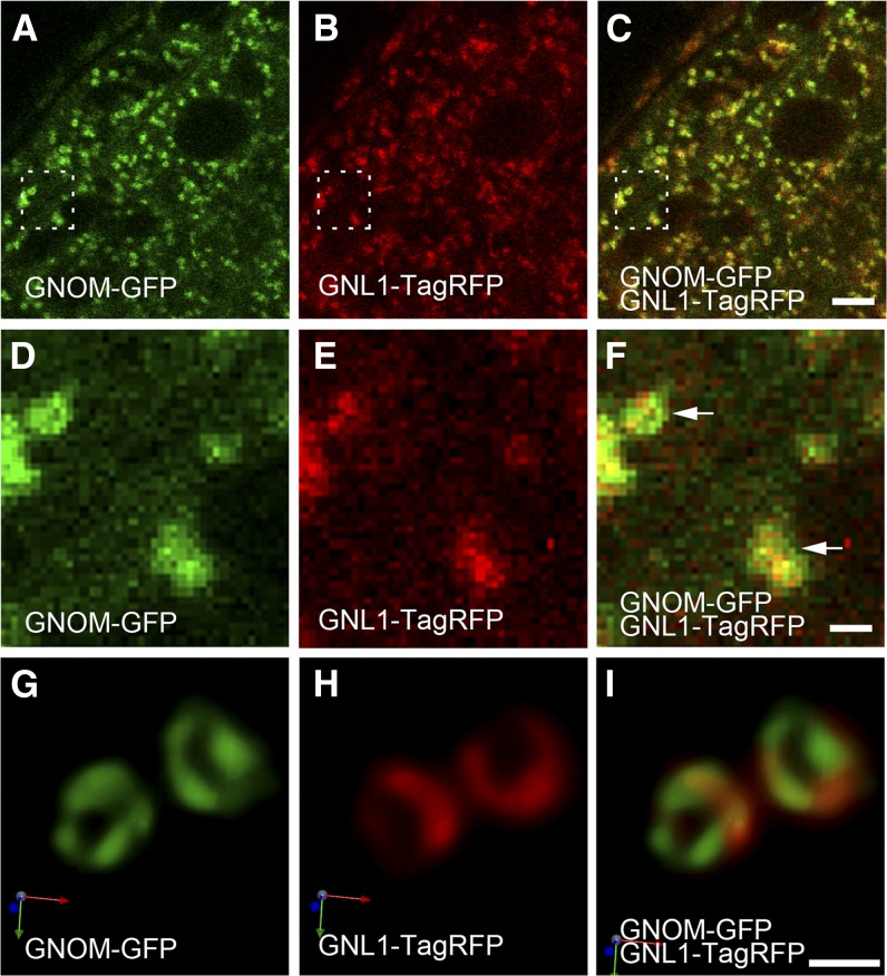
GNOM and GNL1 Localize to Distinct Regions within the Golgi Apparatus.
(A) to (F) CLSM micrographs of Arabidopsis root cells coexpressing GNOM-GFP (green) and GNL1-TagRFP (red). GNOM-GFP (A) and GNL1-TagRFP (B) colocalize to the Golgi apparatus. A merged image is shown in (C). Enlarged views of the dashed frames in (A) to (C) are shown in (D) to (F), respectively. Arrows indicate the apparent distinct localization of GNOM-GFP and GNL1-TagRFP.
(G) to (I) SCLIM micrographs of Arabidopsis root cells that coexpress GNOM-GFP and GNL1-TagRFP. High-resolution confocal analysis identified distinct but partially overlapping subcellular localization of GNOM-GFP (G) and GNL1-TagRFP (H) at the periphery of the Golgi cisternae. A merged image is shown in (I). Images are shown in 3D display and arrows indicate the xyz axis.
Bars = 4 µm for (A) to (C) and 600 nm for (D) to (I).

