Figure 5.
IEM Analysis Identifies GNOM-GFP at the Golgi Apparatus.
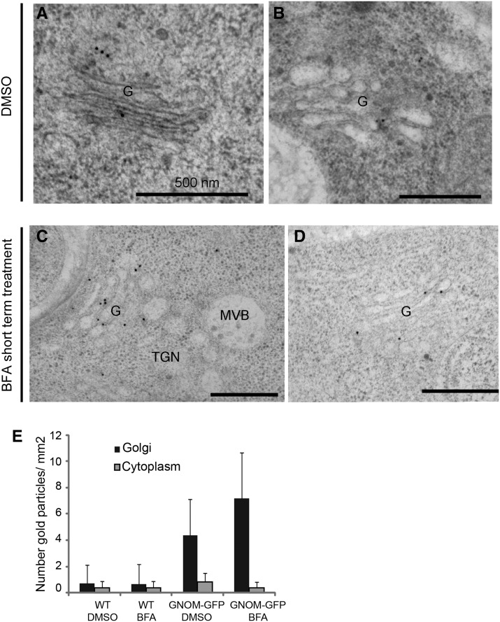
(A) to (D) IEM analysis of GNOM-GFP with anti-GFP antibodies. GNOM-GFP is localized to the Golgi apparatus in DMSO- ([A] and [B]) and BFA-treated ([C] and [D]) cells at the ultrastructural levels.
(E) Quantification analysis of GNOM-GFP localization. Quantification confirmed that GNOM localization at the Golgi apparatus is enhanced after short-term BFA treatment. Error bars indicate sd. Between 20 and 30 Golgi stacks from at least 10 cells in two independent roots were considered for this analysis. Note the weaker or absent detection of GNOM-GFP at the TGN and MVB under either BFA-treated or untreated conditions. The Golgi apparatus is designated as “G” within the figure.
Bars = 500 nm.

