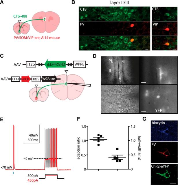
Figure 4.
Long-range GABAergic projection neurons in PFC to NAcc are heterogeneous. A, Experimental design: the retrograde tracer CTb-488 was injected into the NAcc of PV–Cre, SOM–Cre, or VIP-Cre mice crossed with Ai14 mice. B, PV–Cre- and VIP–Cre-labeled neurons in mPFC (red) colabel with the retrograde tracer (green) (scale bars, 15 μm). C, Experimental design of a transynaptic–intersectional GABAergic neuron marker: AAV–mCherry–IRES–WGA::Cre and AAV–Dlxi12b–DIO–ChR2–EYFP viruses were injected into NAcc and mPFC, respectively. D, Images of GABAergic neurons labeled by a transynaptic–intersectional GABAergic marker in layers 2/3 at low and high power (top and bottom rows; scale bars, 60 and 15 μm, respectively). E, Cells labeled by the transynaptic–intersectional tracer were fast spiking. F, The adaption ratios and action potential half-widths for cells labeled in E. G, Transynaptically labeled cells stain for PV. Scale bar, 10 μm. DIC, differential interference contrast; IL, infralimbic cortex; PL, prelimbic cortex.

