
Fig. 3.
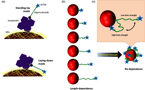
(a) Illustration of two binding modes of biotinylated oligonucleotide to streptavidin-coated quantum dots (QDs). Not to scale. (b and c) Illustration of our hypothesis in this work. At low-ionic strength, oligonucleotide stretches from the QDs, leading to length-dependent FRET. At high-ionic strength, oligonucleotide is more flexible but is prone to bending toward QDs. The separation distance between donor and acceptor becomes smaller, falling well into the FRET range, so that no length-dependent FRET was observed.