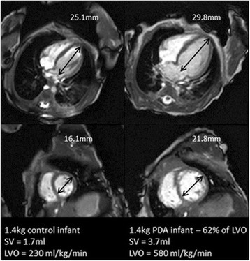
Figure 1.

Comparison of a 4 chamber and short axis view at end diastole in a 1.4 kg control infant (left) and 1.4 kg PDA (right) infant with a shunt volume of 62% of LVO. Apex-base and mid cavity diameter measurements have been included for scale. Figure shows the apparent increase in left ventricular dimensions.
