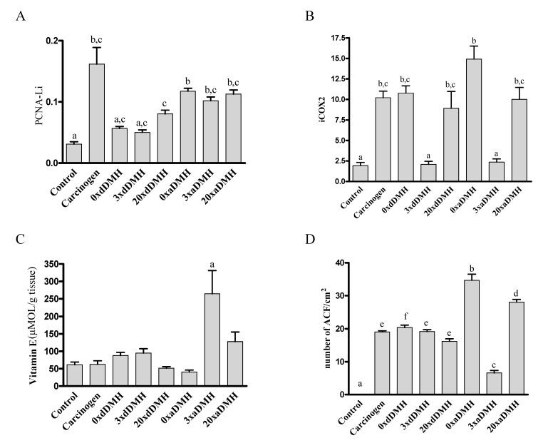
Figure 4.
Colonic expression of PCNA and COX-2 obtained by immunohistochemistry, hepatic content of VE measured by HPLC, and ACF count obtained by H & E. Statistical analysis. (A) PCNA labeling index (PCNA-Li). (B) Cyclooxygenase 2 index (iCOX-2). (C) Hepatic content of VE. (D) Number of ACF. a p < 0.05 compared to Carcinogen, 0×aDMH, 3×aDMH, 20×aDMH; b p < 0.05 compared to Carcinogen, 0×aDMH, 3×aDMH, 20×aDMH and 20×dDMH; c p < 0.05 compared to Carcinogen, 0×dDMH, 0×aDMH, 20×dDMH and 20×aDMH; d p < 0.05 compared to 0xaDMH; e p < 0.05 compared to other groups; f p < 0.05 compared to 0×dDMH.

