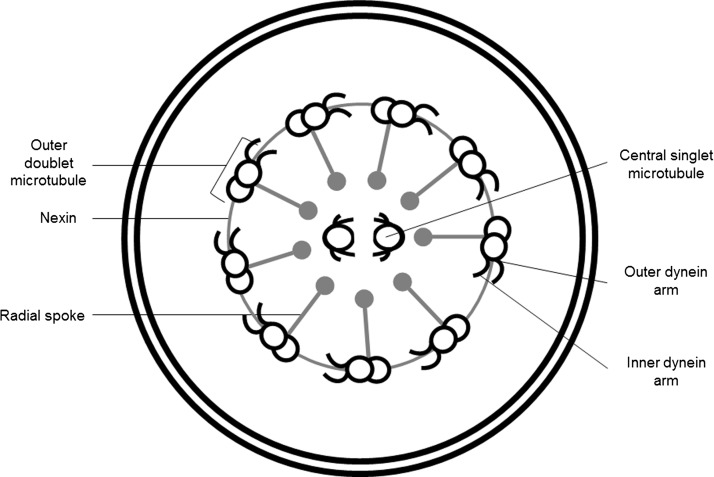
Figure 1.
Cartoon of a respiratory cilium in transverse section as seen by transmission electron microscopy (TEM). Respiratory cilia have a ‘9+2’ arrangement with a central pair of single microtubules running the length of the ciliary axoneme surrounded by nine peripheral microtubule doublets. Nexin and radial spokes maintain the organisation of the axonemal structure. Attached to the peripheral microtubules are inner and outer dynein arms that generate the force for ciliary beating. Abnormalities of the dynein arms affect ciliary beating. HYDIN projections on the central pair of microtubules are not usually seen by routine TEM (Cartoon image provided by Robert L Scott).

