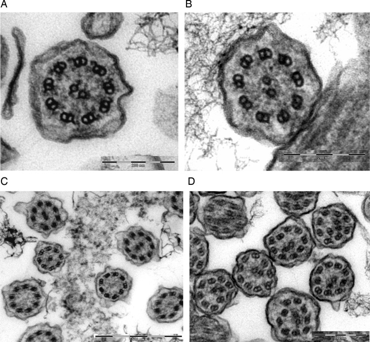
Figure 2.
Transmission EM of a respiratory cilia from (A) a healthy individual. Representative nasal epithelium cilia from patients with primary ciliary dyskinesia (PCD) caused by (B) outer and inner dynein arm defects (C) transposition defect: some cilia demonstrate absence of a central microtubule pair (‘9+0’); in other cilia a peripheral microtubule doublet has crossed to take the central position providing an apparent ‘8+2’ structure (D) microtubular disorganisation. EM images obtained using FEI Tecnai 12 transmission electron microscope (FEI UK Limited, Cambridge, UK) at 80 kV). EM images provided by P Goggin (Primary Ciliary Dyskinesia Centre, University Hospitals Southampton NHS Foundation Trust, Southampton, UK).

