
Figure 1.
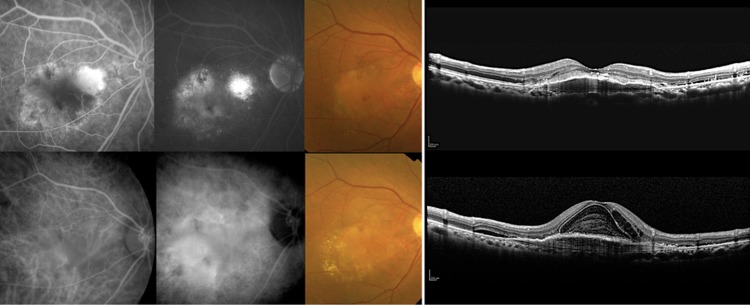
A non-responder as judged by fundus findings with fibrovascular pigment epithelial detachment (PED). This was a 71-year-old man with typical age-related macular degeneration and best corrected visual acuity (BCVA) of 0.3 (logMAR 0.52) at the time of initial intravitreal ranibizumab (IVR). Fluorescein (top row, left two panels, early phase and late phase) and indocyanine green (bottom row, left two panels, early phase and late phase) angiograms were consistent with the findings of the fundus colour photograph and an optical coherence tomography image showing fibrovascular PED before initial IVR (top row, right two panels). After seven IVR injections, the BCVA worsened to 0.15 (logMAR 0.82) at month 12. Although the fibrovascular PED did not change markedly, there was an increase in the subretinal and intraretinal fluid at month 12 (bottom row, right two panels).