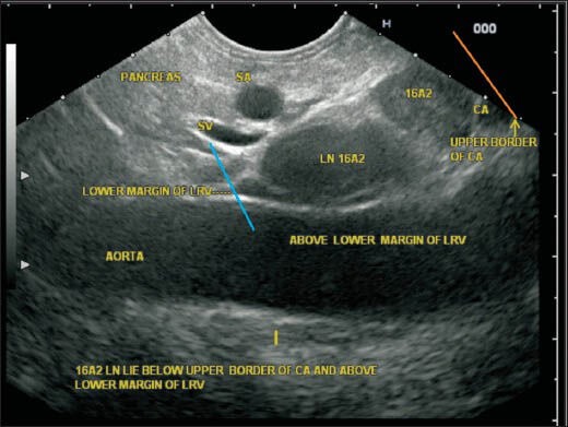
Figure 33.

In this image, the upper and lower boundary of station A2 are seen as the upper border of the celiac artery (CAL) (orange line) and lower border of left renal vein (LRV) (blue line). The lymph node (LN) belonging to station 16A2 lies below the CAL and above the lower margin of LRV anterior to the aorta. Although the LNs lie posterior to the pancreas, the location near the aorta places them in para-aortic group
