Figure 40.

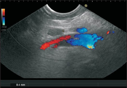
A color Doppler shows the superior mesenteric vein and the lymph node belonging to station 18 from the horizontal part of the duodenum

A color Doppler shows the superior mesenteric vein and the lymph node belonging to station 18 from the horizontal part of the duodenum