
Figure 2.
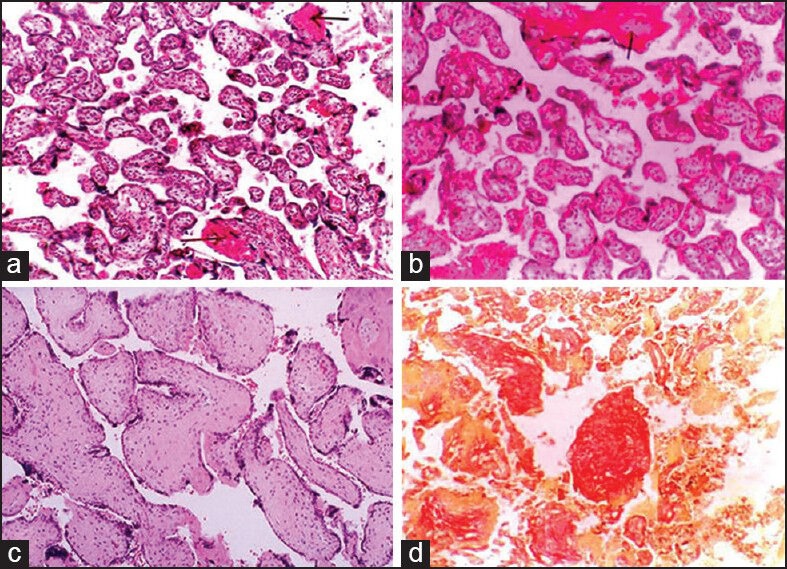
(a) Villi showing areas of fibrinoid necrosis (H and E, ×100), (b) small isolated immature villi among normal villi and area of fibrinoid necrosis (H and E, ×100), (c) villi showing intravillous fibrosis. (H and E, ×400), (d) vilous fibrosis with stromal fibrotic nodule (Van Gieson ×100)